Compare the before and after pictures of different skin problems treated with the therapeutic laser!
Skin Wound
(6a) to (6e) Evolution of a skin wound during laser treatment in a cat at D0, D3, D12, D21 and D60.
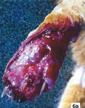
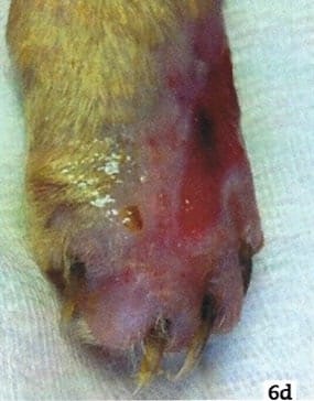
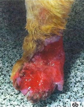
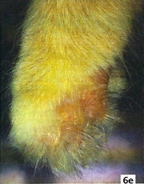
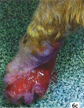
Images of Dr. Susan Kelleher. P.107 Laser Therapy in Veterinary Medicine, Photobiomodulation Edited by Ronald J. Riegel, DVM and John C. Godbold, Jr., DVM
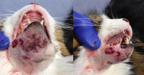
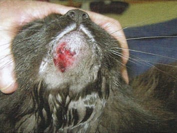
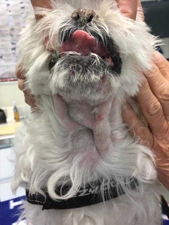
Feline Acnea
February 1st, 2017
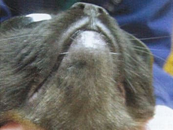
February 2, 2017
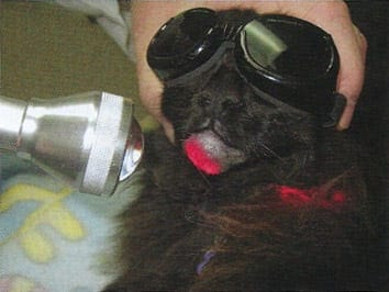
Figure 19.5 shows a case (a) upon presentation, (b) during a laser treatment session (wearing goggles), and (c) on a re-check visit 2 weeks later. This cat is a great example of the rapid positive responses seen.
P.209 Laser Therapy in Veterinary Medicine, Photobiomodulation. Edited by Ronald J. Riegel, DVM and John C. Godbold, Jr., DVM
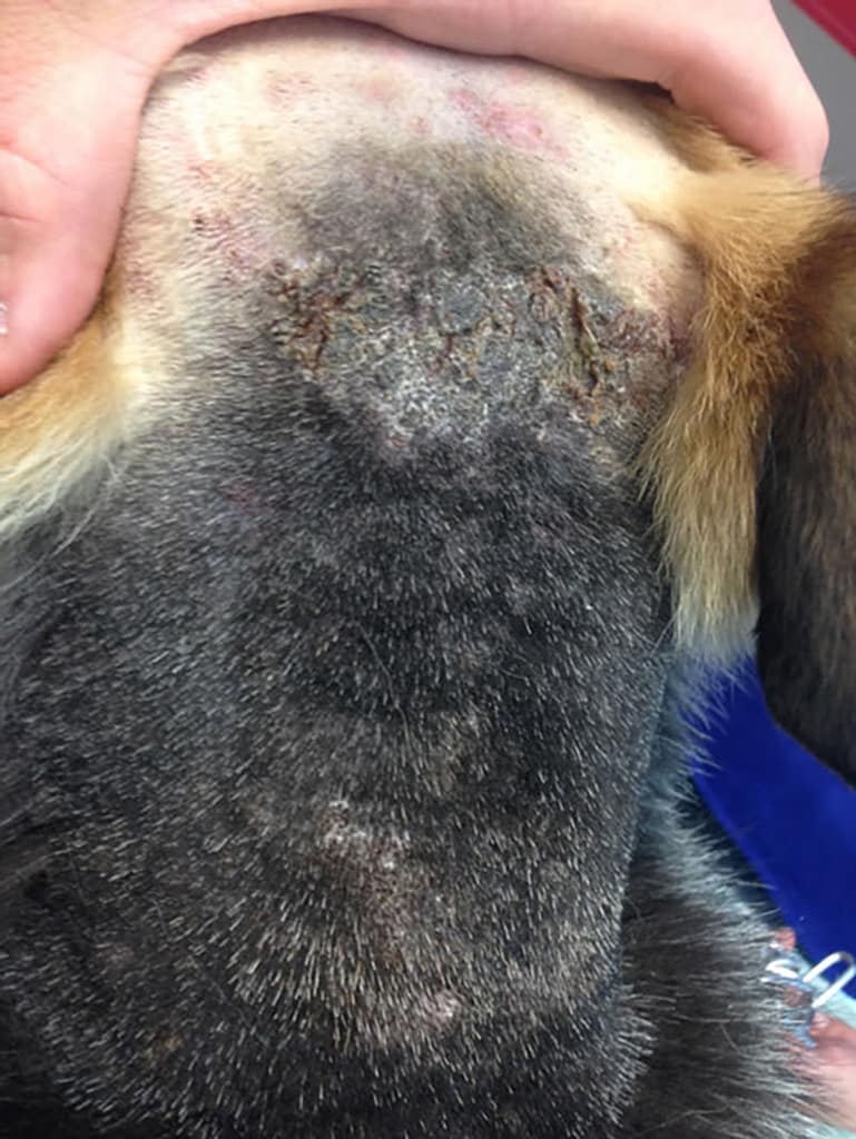
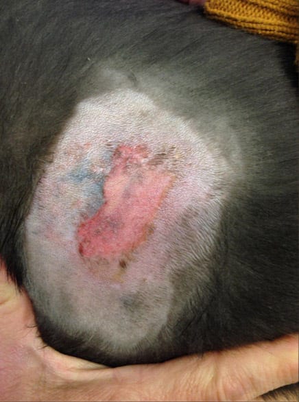
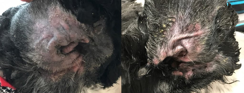
Methicillin-resistant bacterial dermatitis
August 14, 2017
August 17, 2017
Lick Granuloma
“Chronic granulomatous lesion on the front paw of a leopard at a rescue sanctuary. Multiple other therapies, along with environmental enrichment, had been attempted, with no success at reducing continued self-trauma. Laser therapy was instituted (a) and improvement was noted (b).”
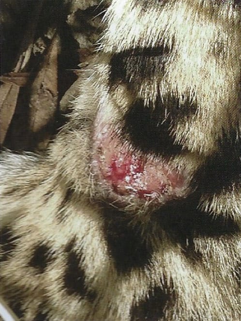
a)
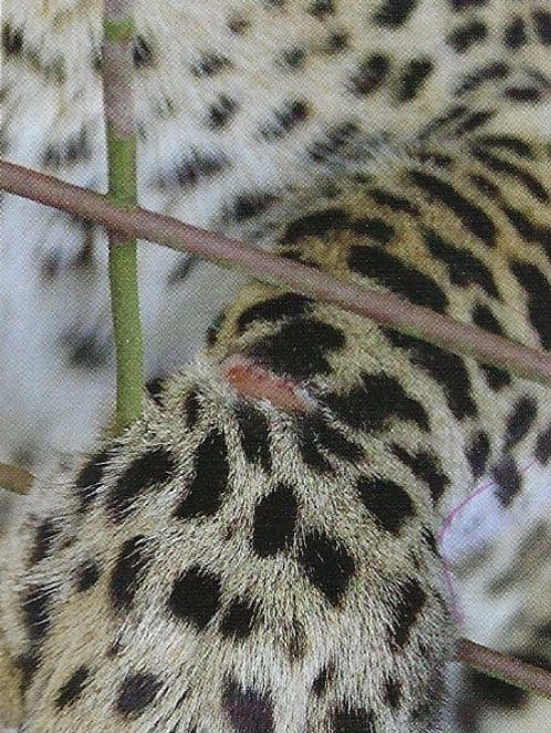
b)
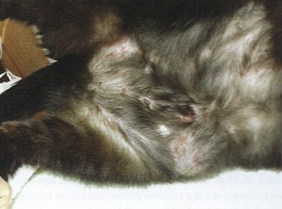
Hot Spot
June 8, 2017
June 10, 2017
Sebaceous gland cyst ruptured
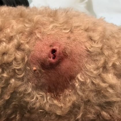
April 12, 2018
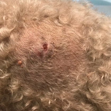
April 17, 2018
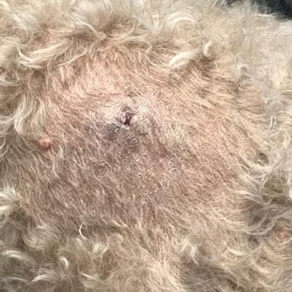
April 21, 2018
April 26, 2018
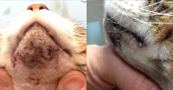
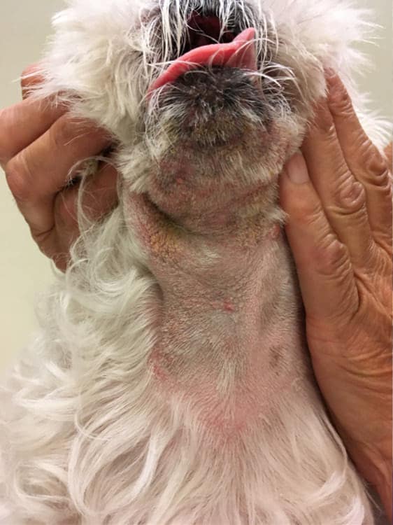
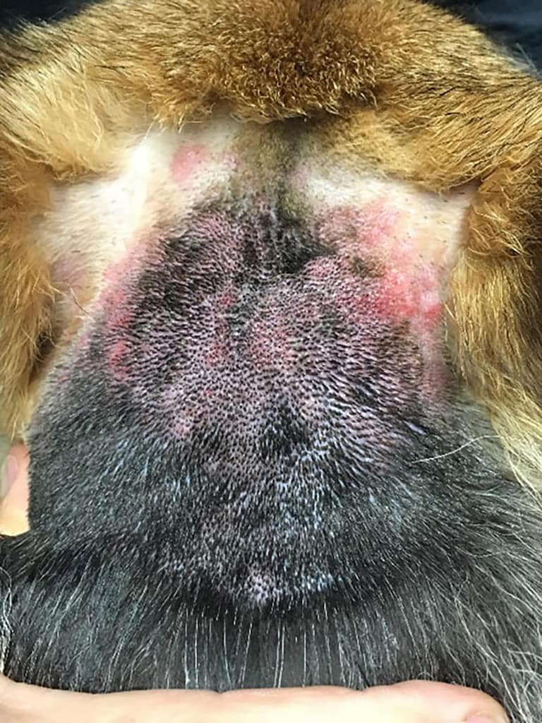
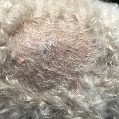
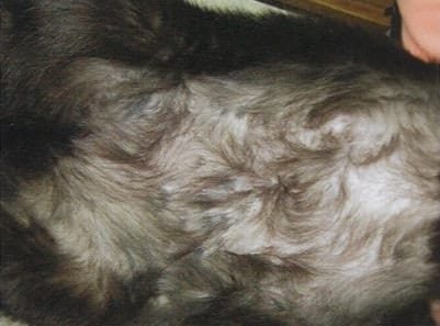
Allergic Dermatitis
A single treatment carried out on 23 May 2017
May 23, 2017
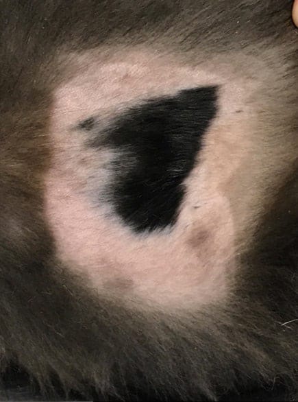
July 10, 2017
Figure 19.6 (a) Self-inflicted excoriation and dermal lesions due to allergic dermatitis on presentation. (b) The same patient without erythema and with significant hait regrowth 3 weeks later, following three laser therapy treatments.
a)
b)
P.107 Laser Therapy in Veterinary Medicine, Photobiomodulation Edited by Ronald J. Riegel, DVM and John C. Godbold, Jr., DVM
Otitis Externa
July 10, 2017
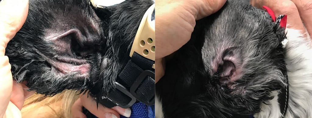
July 17, 2018