Renovating your dentistry room? Planning to buy new equipment? Here are the factors to consider, according to a clinician who has explored several layouts!
X-rays: What You Need to Know Before Buying
Several factors need to be considered when purchasing a new dental X-ray system. If the entire system needs to be replaced, both the X-ray generator and the digital radiography software must be selected. There are many differences between systems. None are perfect, but some may be better suited to your clinic’s specific situation. Here are a few factors to keep in mind.
Choosing the X-ray Generator
Dental X-ray generators have changed slightly in recent years, although the basic functions have remained the same. If you have an older dental X-ray generator, it is likely still compatible with a new system.
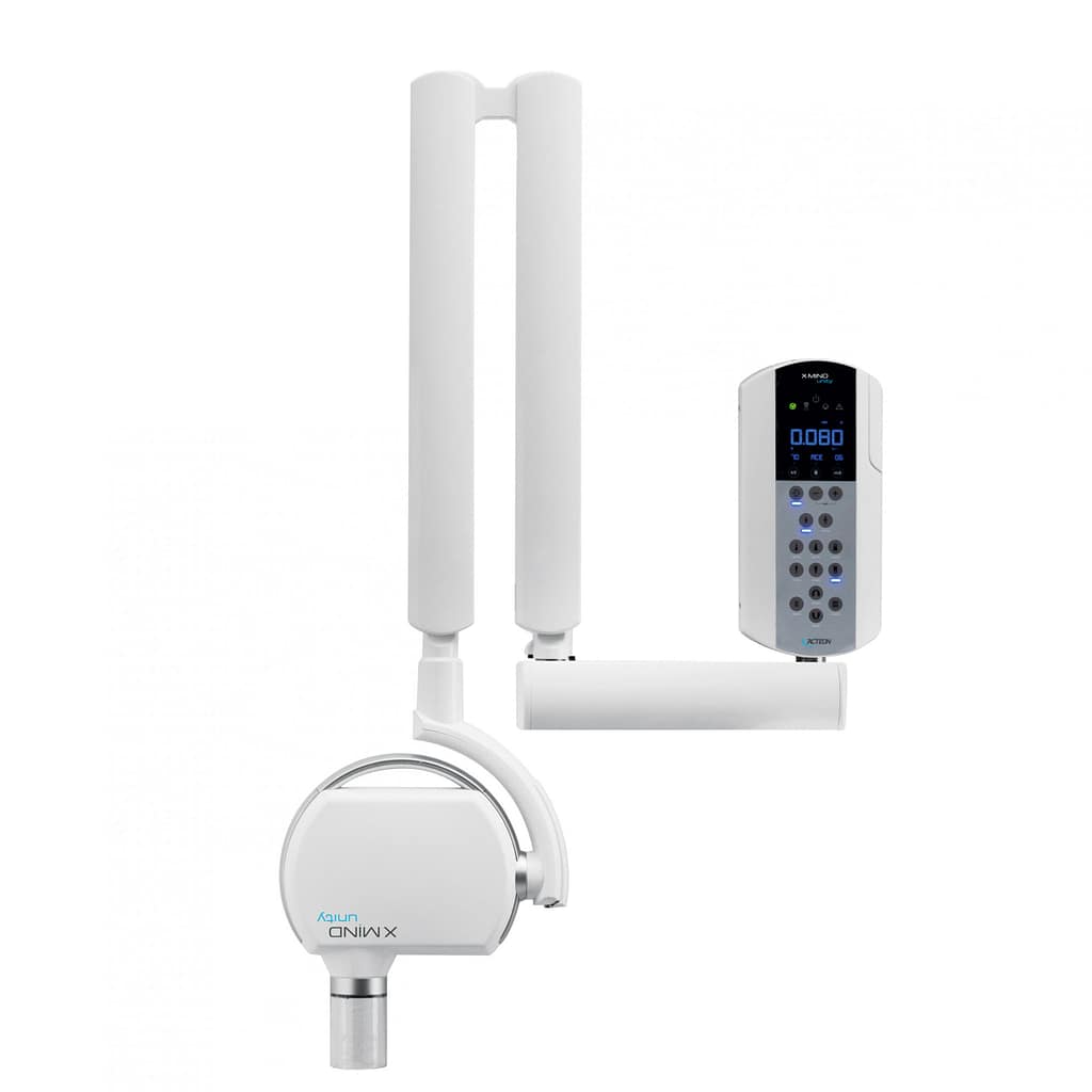
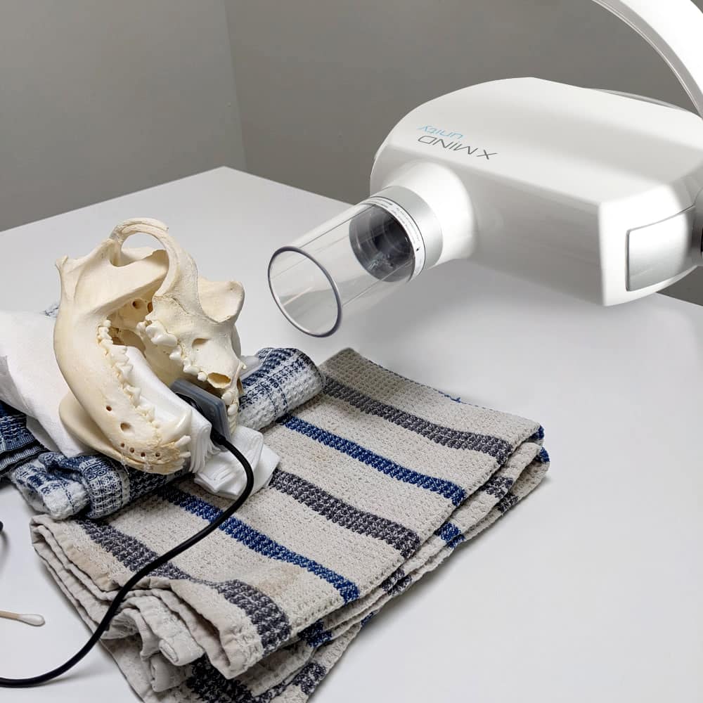
There are mainly three types of dental X-ray generators: wall-mounted, mobile on wheels, and portable (handheld). Wall-mounted generators take up less space in a room. However, once installed, they are fixed permanently. It is important to carefully consider the installation location. The generator must not only reach the end of the table but extend beyond it to ensure all necessary views can be taken.

Think also about larger patients – it is highly inconvenient to have to move the patient during the procedure. You must also take into account potential interference with the surgical light or any other equipment that may be mounted on the ceiling.
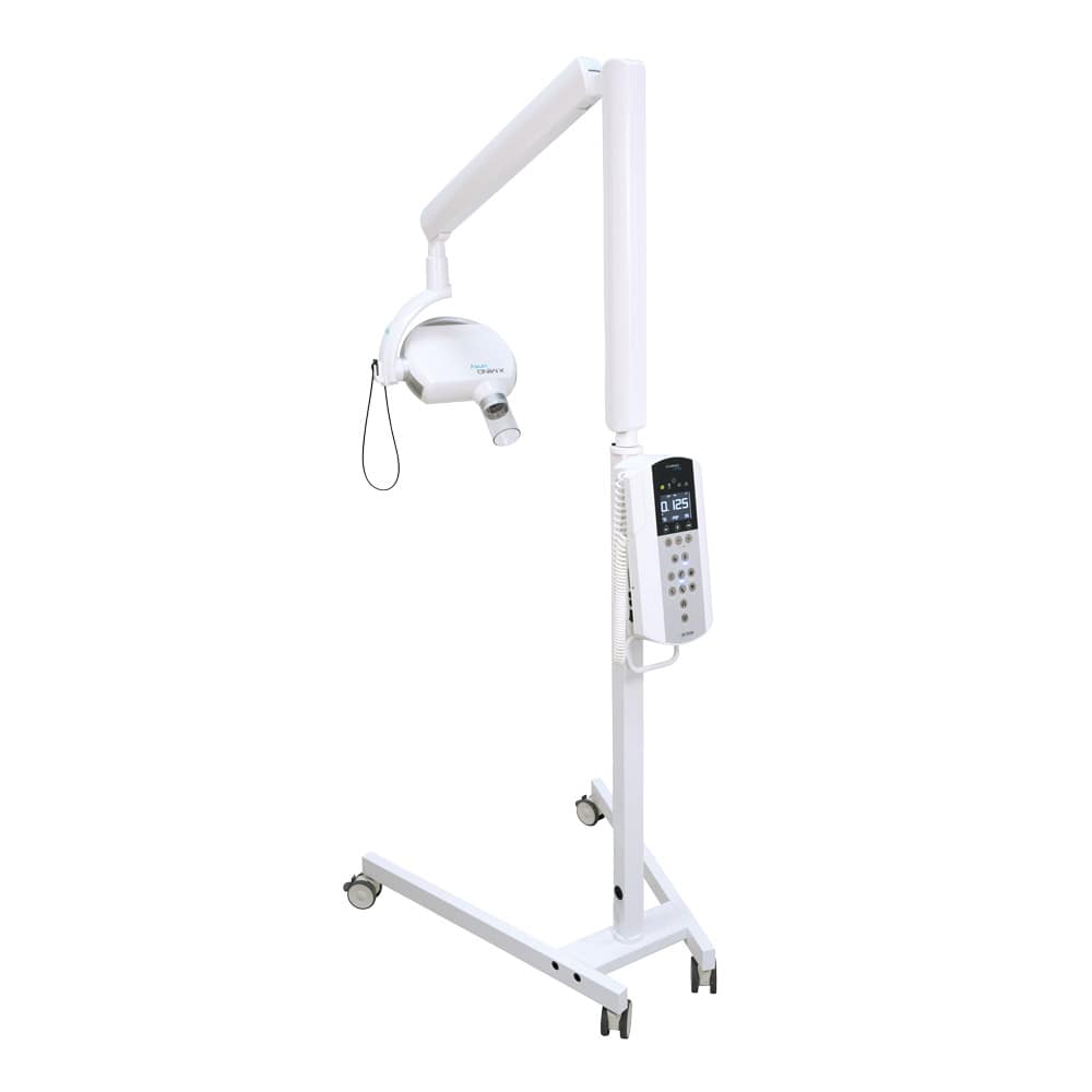
Mobile generators on wheels have the advantage of being stored in another room when not in use. This can be an excellent solution if you perform dental procedures in an area that is sometimes used for other types of treatments. However, these generators are bulky, as they take up considerable floor space, and they can restrict movement or cause accidents if someone trips over them.
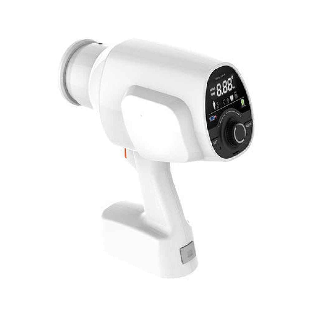
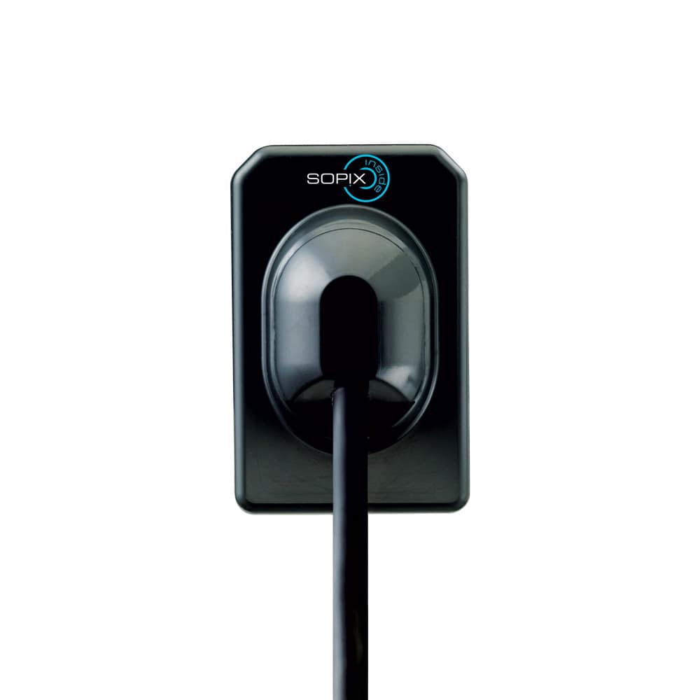
Portable generators are the most compact. However, they require more training to successfully take X-rays from the correct position. Many generators of this type have a digital display that shows the degree of tube angulation to make image capture easier. It is important for the person holding the generator to wear their lead apron and dosimeter.
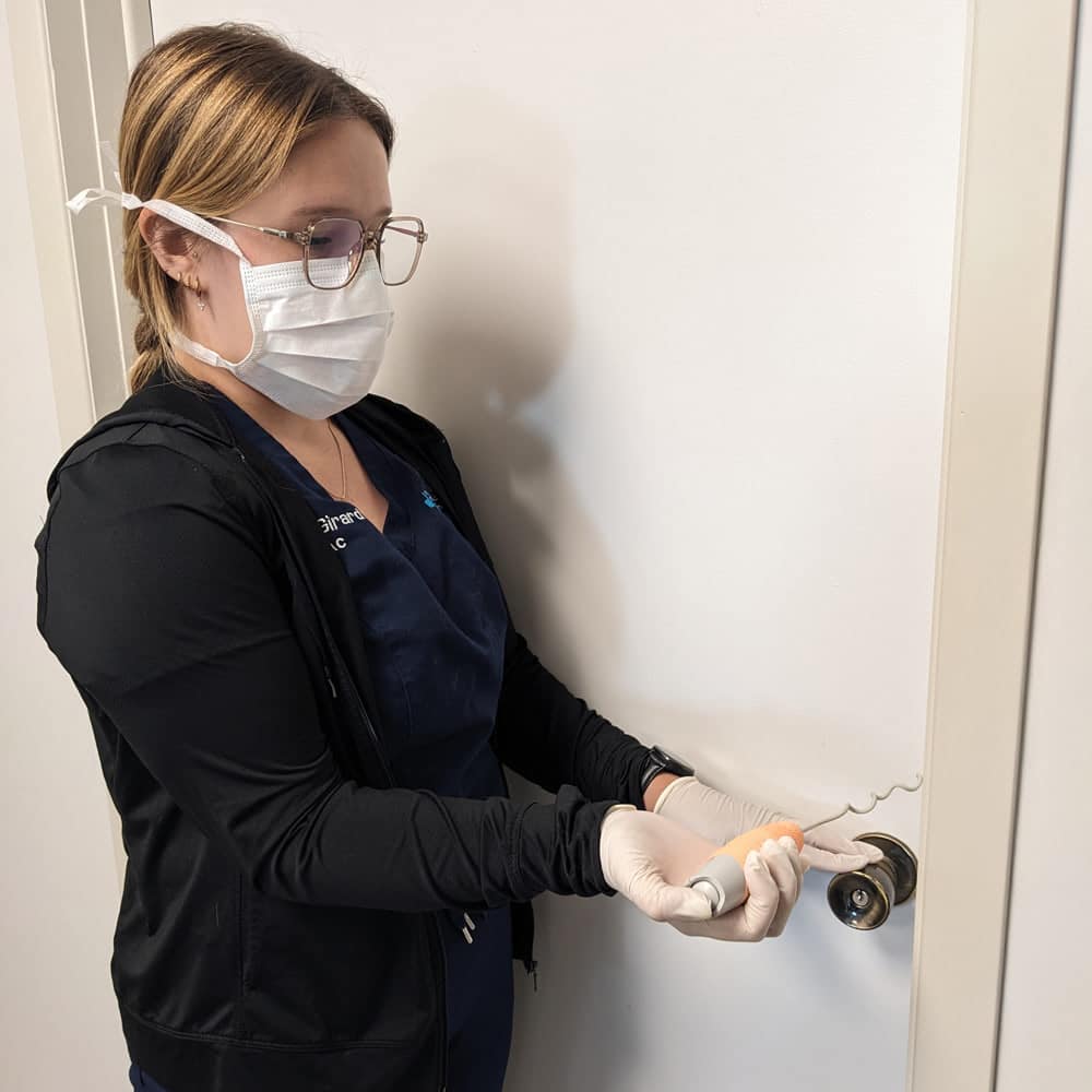
For a wall-mounted or portable generator, several types of triggers are available. Many devices come with a button connected to the unit by a long coiled cord. These triggers are often instantaneous because they use a wired connection, but the cord itself can get in the way of the patient and various attached tubing. It must also be long enough to allow team members to leave the room during image capture.
Some devices come with a wireless remote to trigger image capture. A slight delay is often required before the image is taken. In addition, the remote often ends up misplaced between shots, as we tend to set it down and forget where it is. Much like syringe caps, they seem to vanish from sight the moment we put them down!
The last option is a trigger button mounted on the wall of the dentistry room. This setup offers the benefits of a wired connection and requires team members to leave the room to press the button (as long as it is placed near the door, outside the room), ensuring no one is exposed to radiation. The device is never lost because it’s fixed to the wall, and there’s no cord to get in the way. If your dentistry room is undergoing renovations, it may be worth installing this type of connection. It can also be added later if the clinic has a suspended ceiling.
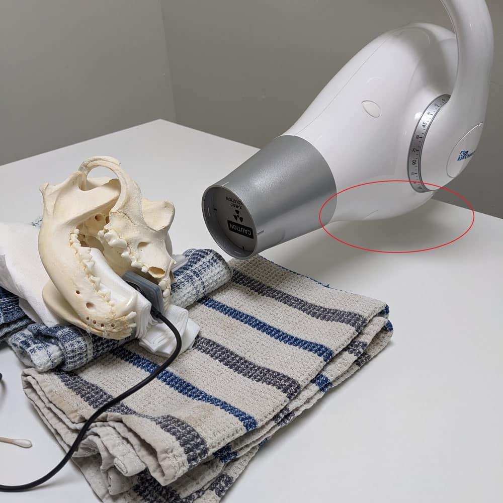
Some generators have been adapted from human dentistry and may include rectangular beam collimation. This feature is not useful in veterinary medicine since the patient is not sitting in a chair but lying on the table. It can become very difficult to aim the sensor with the collimated tube. If your device has this type of collimation, know that it is often just a ring added to the end of the tube, which can be easily removed.
Shape and Quality of the Generator: Details That Matter
One factor to consider is the shape of the generator. Again, since these generators are often designed for human dentistry, they can be mounted either horizontally or vertically. Vertical generators often interfere with the procedure table and should be avoided.
Vertical Generator
Horizontal Generator
Finally, the quality of the generator should be considered. A smaller focal spot (around 0.6 to 1 mm) will produce clearer images. The price is usually set accordingly. Keep in mind that in general practice, a slightly larger focal spot will likely not affect your interpretations.
Choosing the Radiography System
Once the generator is chosen, you need to select a radiography system. There are two main types of systems: direct and indirect. Here are their main characteristics.
DR (Digital Radiography): The Direct System
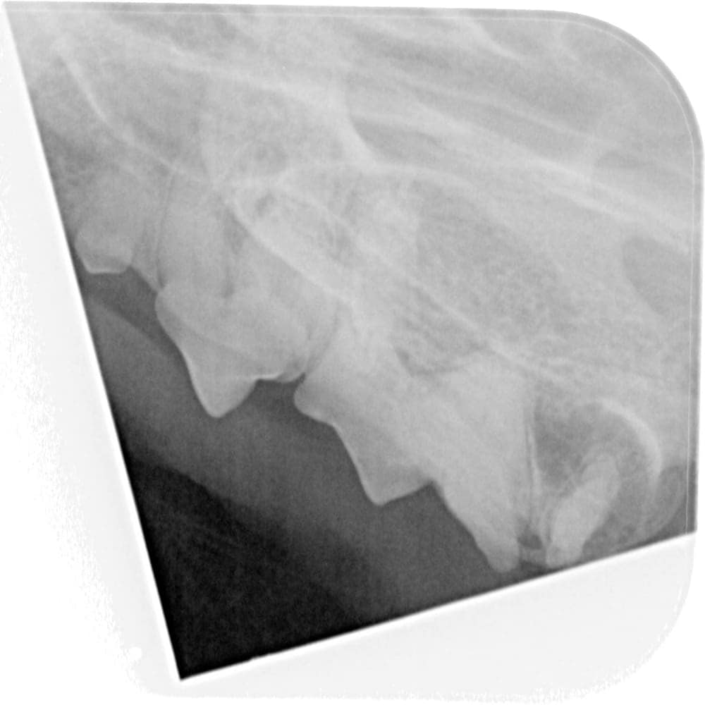
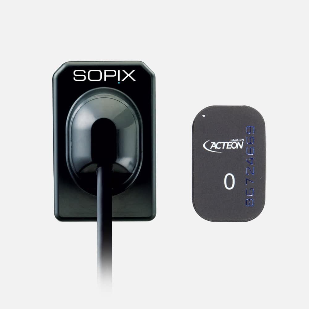
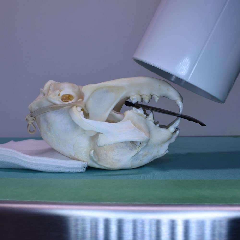
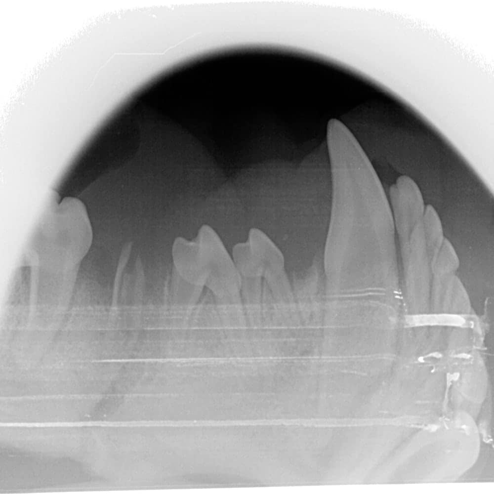
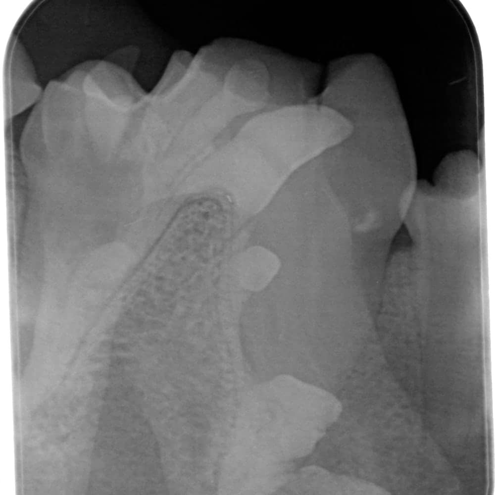
Direct systems, or DR (Digital Radiography), are the most commonly used systems. Films are replaced by sensors, which are available in several sizes -commonly sizes 0, 1, 2, and 4. The sensors are several millimeters thick and are connected to the device (the generator or computer) by a cable.
Sometimes the photosensitive cells don’t extend all the way to the edge of the plastic casing, so the actual sensor is smaller. The sensors are expensive and represent an investment of several thousand dollars; clinics generally choose only one size 2 sensor to work with. It is important to protect the sensor by avoiding dropping it or having a lightly sedated patient bite it. Radiographs under sedation are avoided only with this type of device. The cable can also be a source of damage if it becomes bent or twisted. The main advantage of this system lies in the fact that images are available instantly on the screen at the time of capture. Moreover, since the sensor is always in the animal’s mouth, it is easier to correct a bad shot by slightly moving the sensor and immediately retaking the image. These systems are compact because the only device needed is the sensor itself, connected directly to the computer or generator.
CR (Computed Radiography): The Indirect System
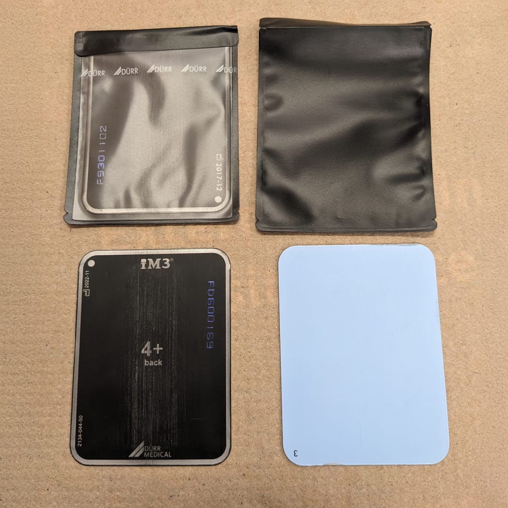
Indirect systems (indirect digital, computed radiography), or CR, use photostimulable phosphor plates to capture the image. These plates are available in several sizes (0, 1, 2, 3, 4, 5) and are about the thickness of a playing card.
These plates must be placed in protective plastic sleeves. Manufacturers recommend using a new sleeve for each EXPOSURE, although they can be reused a few times. Some brands of sleeves need to be torn in the center to be developed, making them unusable a second time. Although phosphor plates are less expensive than digital sensors (about $150 for a size 2 plate and $600 for a size 4), the cost of the sleeves must be considered. They can cost around $1 each, which increases operating expenses. On the other hand, it is less problematic if a patient bites the plate due to its more affordable price.
Phosphor plates can scratch more easily and delaminate if not cleaned with 99% alcohol. They must therefore be replaced after a few years, unlike digital sensors. The variety of sizes offered by this system is its main advantage: size 0 plates easily fit into the mouth of a 1 kg patient or a small cat. Size 4 plates are very useful for taking fewer shots on a large dog and provide a better overall view.

Sometimes, size 4 plates are used to X-ray small companion mammals – this type of system could be advantageous if you treat these kinds of patients in your clinic. Size 5 plates are sold by representatives but, in my humble opinion, offer little benefit in veterinary medicine. Two plates of sizes 0, 2, and 4 make a good kit for all patients.
Phosphor plates must be developed once exposed. This requires a digital developer placed on a countertop in the dentistry room. Developers can vary in size but consider approximately the size of a toaster. Since the plates need to be developed, there is a slight delay in image processing. Development takes about 30 seconds, during which you can take the next shot with a second plate. However, if a mistake was made during the image capture, it may be more difficult to correct the situation because the plate will have already been removed from the animal’s mouth. More artifacts are possible when using phosphor plates. For example, blood on the cassette will be visible during development.
A double exposure can occur if you forget to develop the plate. The main disadvantage is that the plate could be placed upside down in the patient. Indeed, the blue side of the plate should face the X-ray generator. If the plate is placed upside down, the resulting image will be a mirror image of reality. It would be unfortunate to extract a tooth on the wrong side if you don’t realize this mistake. Indirect systems are often slightly more expensive to purchase than direct systems – unless a sensor breaks!
Choosing the Software: Ergonomics and Backup
Once the system is chosen, you can explore various brands to see if you like their software. The software should allow you to find a patient, compare X-rays side by side, and easily export images. Most representatives can lend you a device for a few days so you can try it out.
Most radiography software will recommend having a computer dedicated to this function. If your computer is not connected to a network (PACS), be sure to regularly back up your data to avoid losing it.
Room Layout: Think Ergonomics and Efficiency
Please carefully consider the room’s ergonomics if your dentistry room is being renovated. Because of X-rays, the team needs to enter and exit the room frequently, which is unique to dentistry’s requirements. Think about placing the exit near the patient, taking into account the anesthetic machine’s tubing without blocking access to the door. The door could be wider than a standard door, and two doors are often very useful when the clinic’s layout allows it.
Choosing the Right Dentistry Table
Another addition to your setup is the dentistry table. Again, several factors need to be considered, and there is no one-size-fits-all solution. It is not strictly necessary to have a table with a built-in sink to perform dental procedures. Although water is produced during scaling, the amount is minimal and can be absorbed by a towel. Obviously, the sink can have other uses. Tables with a drain at one end are an excellent compromise.
Most of these tables are height-adjustable, which greatly increases user comfort. Adjustable tables are ideal in dentistry since patient sizes vary. They also allow the surgeon to work standing or sitting, depending on preference.
It can be helpful to consider the location of the foot pedal on this type of table. Adjustable tables generally have a pedal on the side because they are designed for general surgery. In dentistry, the surgeon is usually positioned at the end of the table, making the pedal inaccessible. Some tables have the pedal at the front.
Other tables feature a rounded end. These tables are ideal for dental procedures because they allow the surgeon to slightly turn around the patient without the corner of the table pushing them away from the patient.
Workstation Ergonomics
The table should be at a height that allows your elbows to be bent at 90 degrees close to your body to avoid fatigue. Loupes and headlamps also greatly improve posture. Loupes equipped with a light move with the user and don’t need to be constantly adjusted.
An overhead surgical light isn’t strictly necessary if you have a headlamp. Loupes are also useful during consultations because they allow a thorough oral exam with excellent lighting. Lighting in a consultation room usually comes from the ceiling, and when you lift the patient’s lips, shadows are inevitably cast. Additionally, it shows the client that an oral exam is important, which can set the stage for the discussion that follows.
Loupes can also be useful during general surgery. Some people need time to get used to loupes, while others wear them comfortably from the first use. A 2.5x magnification is sufficient for a first pair of loupes.
It’s generally a good idea to choose a company with a local representative who can adjust the loupes as needed and perform simple repairs quickly in case of damage. Most companies offer low-cost prescription changes for loupes, so they typically last several years.
Other Essential Accessories for Comfort and Efficiency
An ergonomic bench, or at least an adjustable one, is also a good investment. Dental procedures can be long, and the surgeon’s comfort improves tolerance for these types of procedures!
The instrument table should be positioned so the surgeon doesn’t have to turn to reach the instruments. They should also be within easy reach.
It’s also helpful to place the computer screen (or a screen that only displays the computer’s images) facing the surgery table so the veterinarian can view X-rays during extractions without having to turn again.

Written by
Dr. Josée Marcoux
It was during a return to her studies that Dr. Marcoux decided to pursue her dream of becoming a veterinarian, earning her degree from the Faculty of Veterinary Medicine at the Université de Montréal in 2016.
Her profile naturally drew her toward specialty medicine, as early as her second year of studies she began her path in veterinary dentistry… [Read More]